Kortikale Atrophie Frontoparietal
Atrophy also destroys the connections that help the cells communicate. Though often no identifiable cause is found.

Pathologie Druckversion Wikibooks Sammlung Freier Lehr Sach
This part of the brain is responsible for a number of very important processes and as a result changes to its shape and structure can cause a variety of problems.
Kortikale atrophie frontoparietal. Mri shows frontoparietal cortical atrophy predominantly in the paracentral region. The most common way i see the phrase used and in which i use the phrase myself in daily practice is when referring to diffuse cortical volume loss atrophy related to the normal aging process which is essentially normal as it is an expected change as patients enter their 60s. Hyperintensities on t2 weighted mr may be seen in the motor region cortex and subcortical white matter.
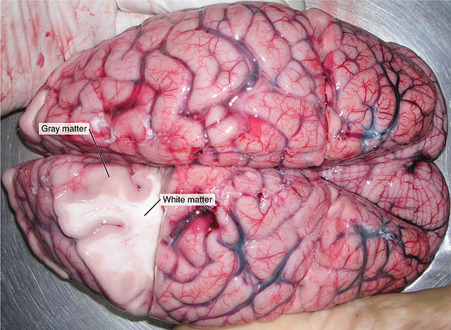
In the preclinical ad group not only frontoparietal but also temporal and cingulate regions and the amygdala diminished in size. Frontal lobe atrophy is a reduction in size of the frontal lobe the foremost area of the brain. Answer cortical brain related atrophy means wasting away and decrease in size of gray matter of brain.
Shrinkage of temporal and cingulate regions greatly accelerated. Brain atrophy is shrinking of the brain caused by the loss of its cells called neurons. Cerebral atrophy is the morphological presentation of brain parenchymal volume loss that is frequently seen on cross sectional imaging.
Brain atrophy or cerebral atrophy is the loss of brain cells called neurons. Rather than being a primary diagnosis it is the common endpoint for range disease processes that affect the central nervous system. Symptoms of significant brain atrophy include progressive cognitive impairment involving multiple cognitive functions otherwise known as dementia seizures and aphasia which is the disruption in the understanding or production of language or both.
Diffuse means the wasting is generalizedgeneralized anxiety disorder not confined to one particular area. The cerebral atrophy is a neurological condition characterized by the progressive death of brain neurons. Cortical atrophy in alzheimers patients brain.
This pathology is characterized by affecting specific regions of the brain which is why it can be divided between cortical atrophy and subcortical atrophy. The mild atrophy in these groups contrasted with the symptomatic ad patients where atrophy reached its peak. Temporoparietal atrophy may therefore provide a useful marker of the presence of ad pathology even in subjects with atypical clinical presentations especially in the context of relative sparing of the hippocampus.
It can be a result of many different diseases that damage the brain including stroke and alzheimers disease. The suggestive clinical signs include asymmetrical motor disorders predominantly in the upper limbs sensory abnormalities and apraxia.

Posterior Cortical Atrophy Radiology Reference Article
Http Www Mri Roentgen Ch Pdf Patienteninformation Mri Demenz Pdf
Https Www Thieme Connect Com Products Ejournals Pdf 10 1055 A 1073 2101 Pdf

Visual Processing Speed Is Slowed In Pca And Predicts

Qualitative Assessment Of Brain Morphology In Acute And Chronic
Https Pdfs Semanticscholar Org 7e60 4594f3330a1ef0c3402f2063740a76295042 Pdf

Telencephalon Anatomie Und Funktion Des Grosshirns

A Review Of Magnetic Resonance Imaging Studies Of Brain
Https Pdfs Semanticscholar Org 7e60 4594f3330a1ef0c3402f2063740a76295042 Pdf