Cortical Desmoid Knee
Desmoid tumors that grow can extend to involve nearby tissues and organs causing signs symptoms and complications. Another term for desmoid tumors is aggressive fibromatosis.
Bone Tumor Mimickers A Pictorial Essay Mhuircheartaigh Jn Lin Yc

Desmoid tumors most often occur in the abdomen arms and legs.

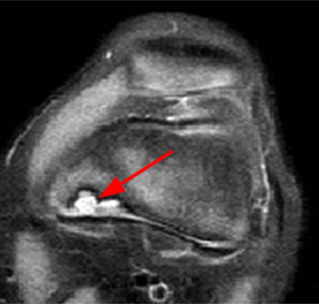
Cortical desmoid knee. It often needs no follow up examination in asymptomatic patients. Cortical desmoid is one of the most common incidental osseous findings on conventional radiographs and mri of the knee. Cortical desmoids are classically seen at the posteromedial aspect of the distal femur.
Distal femoral metaphyseal irregularity cortical desmoid is an irregular cortical margin with an associated lucency found in the posteromedial aspect of the distal femoral metaphysis. They can be bilateral in approximately one third of cases. Nonossifying fibromas nofs are the most common benign bone tumor in children.
Malignancy needs however to be ruled out. Note the cortical irregularity of the distal posterior femur on the left side yellow arrow with no associated bone destruction or soft tissue mass. It is thought to be an avulsion off the medial supracondylar ridge of the distal femur.
Treatment may involve surgery to remove the desmoid tumor when possible. Cortical desmoid is one of the most common incidental osseous findings on conventional radiographs and mri of the knee. They are more common on the left and are bilateral in up to 35 of cases.
Desmoid comes from the greek word desmos which means tendon or band like. Desmoid tumors are usually considered benign not cancer because they rarely spread to different parts of your body. Cortical desmoids are one of the most frequent incidental findings on x rays and a common finding in mri.
Nofs are often discovered by chance when a patient requires x rays for another reason such as a knee injury. It often needs no follow up examination in asymptomatic patients. The age and the incidental detection asymptomatic of the lesion are also typical.
Although frequently bilateral the right knee in this patient is normal white arrow and demonstrates the normal appearance of the cortex. The characteristic location of the cortical desmoid on the dorsomedial aspect of the distal femur the attachment of the adductor magnus or medial head of gastrocnemius is also well illustrated in this patient. It is estimated that 30 to 40 of people under the age of 20 have an nof although few will have any symptoms.
They are seen in up to 5 of young women and in up to 10 of young men. Occasionally similar lesions have been described involving the humerus medially at the insertion of the pectoralis major muscle or laterally at the insertion of the deltoid 9.

Bone Tumor Mimics Avoiding Misdiagnosis Sciencedirect

A D Case 2 A Anteroposterior View Of The Right Knee Demonstrates
Developmental Variants Radsource
Cortical Avulsive Injury Cortical Desmoid In A 12 Year Old Boy


Cortical Desmoid Radiology Reference Article Radiopaedia Org
2
Non Ossifying Fibroma Pathology Orthobullets
Https Www Ajronline Org Doi Pdf 10 2214 Ajr 10 4815

Musculoskeletal Tumors Diagnostic Imaging Of Infants And