Cortical Desmoid
It is thought to be an avulsion off the medial supracondylar ridge of the distal femur. Cortical desmoid also called tug lesion or periosteal desmoid is an irregularity of the distal femoral cortex caused by repetitive stress at the attachment of the adductor magnus aponeurosis.
Cortical Desmoid Radiology Case Radiopaedia Org
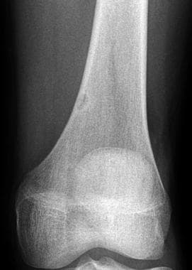
Contrast this with the appearance of the typical non ossifying fibroma on the dorsolateral aspect of the distal femur.
Cortical desmoid. It can press against blood vessels and nerves and cause pain a limp or problems using your legs feet arms or hands. Although it is rarely described in the orthopedic and sports medicine literature knowledge is of high clinical relevance. To confirm the diagnosis some or all of the tumor might be removed with surgery and then tested in a lab.
Desmoid tumors are benign which means they are not cancer. Epidemiology it typically presents in adolescents 10 15 years of age. It is most commonly seen in adolescents and is usually asymptomatic.
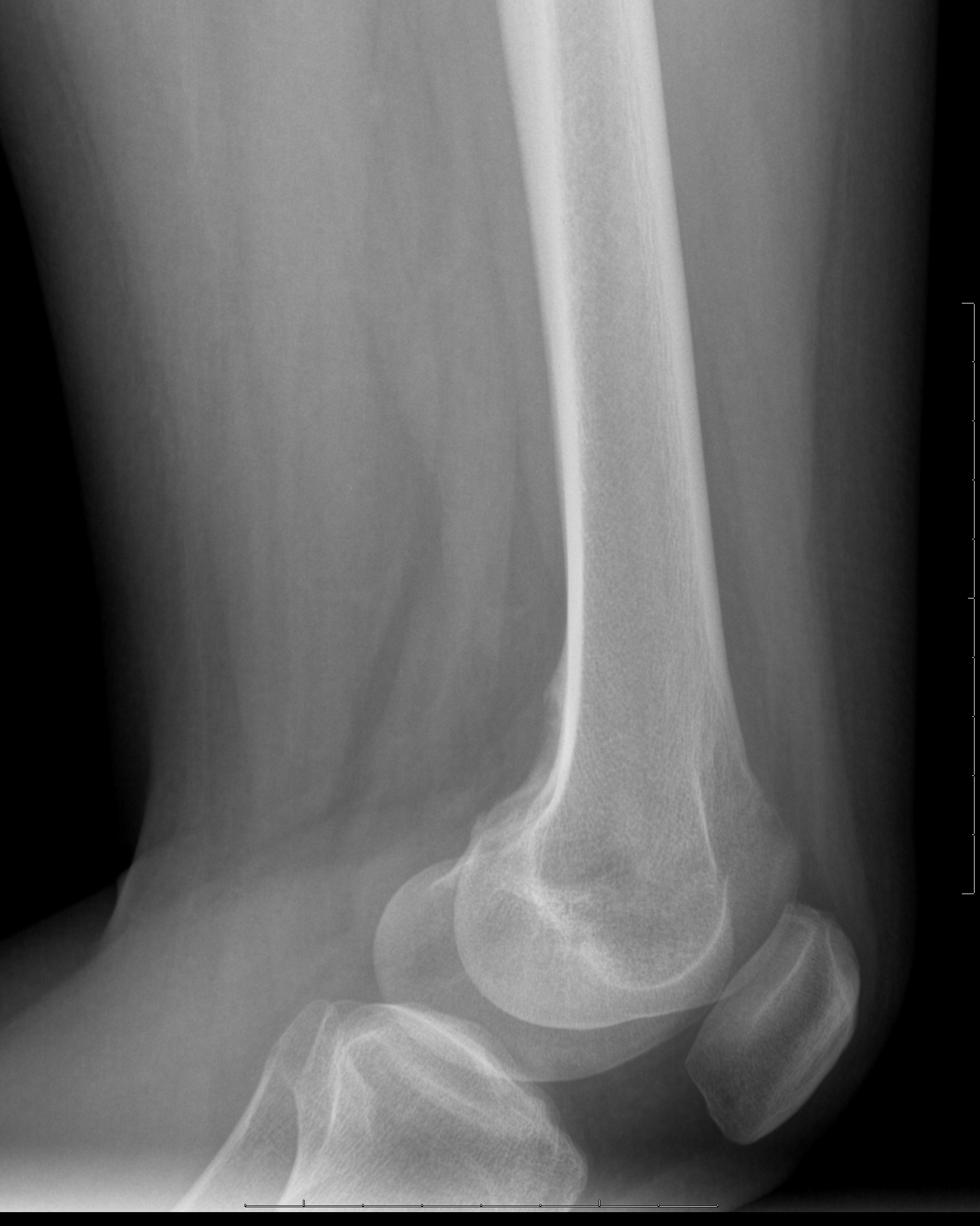
Cortical desmoid is a misnomer as this lesion does not histologically correlate to true desmoid tumors. Cortical desmoid on the dorsomedial aspect of femur right side of image. To diagnose desmoid tumors doctors use imaging tests like ct and mri to create pictures of the tumor.
Signs of a desmoid tumor depend on where it is. A genetic syndrome that causes many colon polyps. Desmoid tumors are often found in the abdomen as well as the shoulders upper arms and thighs.
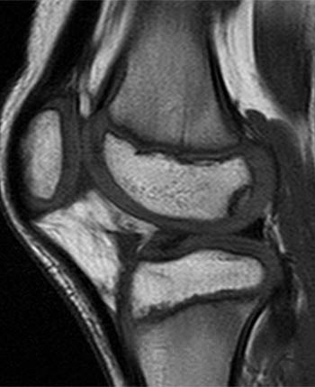
Desmoid tumors tend to occur in younger adults in their 20s and 30s. Axial t2 of the left knee. Largely extra cortical lesion with cortical irregularity on the dorso medial aspect of the distal femur.
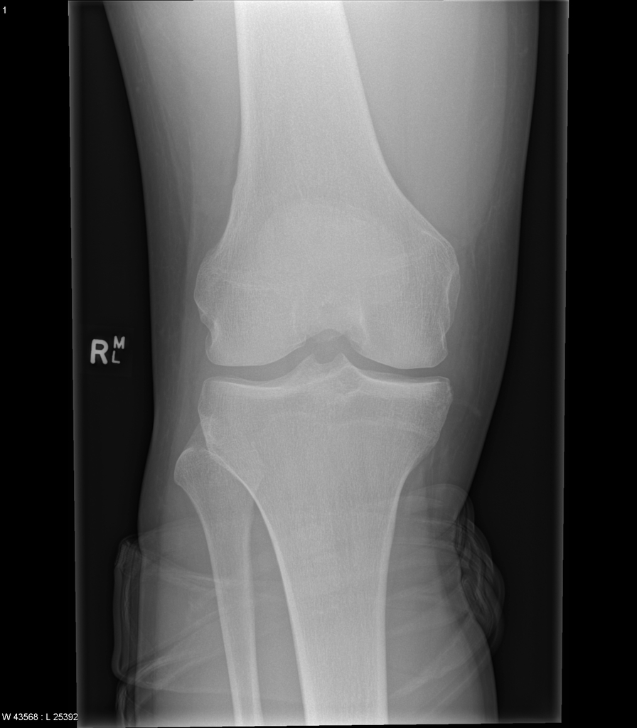
A desmoid tumor can occur anywhere in the body since connective tissue is found everywhere in your body. Distal femoral metaphyseal irregularity cortical desmoid is an irregular cortical margin with an associated lucency found in the posteromedial aspect of the distal femoral metaphysis. Although frequently bilateral the right knee in this patient is normal white arrow and demonstrates the normal appearance of the cortex.
People with familial adenomatous polyposis fap have an increased risk of desmoid tumors. Cortical desmoid represents a benign cortical irregularity typically located at the posterior medial condyle of the femur. If its close to the surface of your skin you may have a painless or slightly painful lump.
This tumor is rare in children and older people. Desmoid tumors are also known as aggressive fibromatosis or desmoid type fibromatosis. Note the cortical irregularity of the distal posterior femur on the left side yellow arrow with no associated bone destruction or soft tissue mass.
If its in your abdomen it may be more aggressive. It is a benign and self limiting lesion.
Cortical Desmoid Image Radiopaedia Org
Https Www Ajronline Org Doi Pdf 10 2214 Ajr 10 4815
Https Encrypted Tbn0 Gstatic Com Images Q Tbn 3aand9gcskfqzwi9zzafyc7laovdk2ty9cpymrbfghyypgexum8ba8xqnjmgd97a
Https Encrypted Tbn0 Gstatic Com Images Q Tbn 3aand9gcq3hozzj6wqv55zxi7olopffdmbxgsebdabic3qxvjzg Jhqsln9rgtyc
Developmental Variants Radsource
Bone Tumor Mimickers A Pictorial Essay Mhuircheartaigh Jn Lin Yc


Cortical Desmoid Fifa Medical Platform
Https Www Clinicalimaging Org Article S0899 7071 18 30011 1 Pdf
Imaging In Fibrous Cortical Defect And Nonossifying Fibroma